Request form
The US version of the website is under construction.
Important:
SIGNUS Medizintechnik GmbH only provides general information about spinal conditions. Please direct specific questions about your situation to your doctor. We cannot accept liability for incorrect indications and/or treatment and their consequences.
Anatomy of the spinal column
Even the words used in the term ‘spinal’ and ‘column’ indicate the special feature of this ‘organ’. ‘Spinal’ represents a central feature, the main source of strength of something. The ‘column’ indicates stability and reliability.
All this describes a complex system that is made up of very different components. To be able to understand the whole system, first the individual parts have to be understood.
So that you can get a better overview of your complex system ‒ the spinal column ‒ we would like to give you an in-depth look into the anatomy and interaction between the individual components of the spinal column.


The Spinal Column
The spine is made up of vertebrae that are joined together by several joints. Each joint allows small movements and all the joints working together make the spine very flexible.
The spinal column is divided into the upper (cervical), middle (thoracic) and lower (lumbar) back. The tailbone is a triangular bony structure made up of four to five fused bones that joins to the lumbar spine. The discs sit between the vertebrae. The iliac bone and the tailbone together form the sacroiliac joint (SIJ) which connects the spinal column to the pelvis. If you look at the spine from the side, you can see a double-S shaped curve that protects the spine from shocks and helps it to best manage the stresses and requirements of everyday life. This shape is produced by the different curves in the individual sections of the spine. The upper and lower back curve forwards and create lordosis while the middle back and the sacrum curve backwards and create kyphosis.

The perfectly coordinated interaction between the bony parts, joints, connective tissue, ligaments and discs means that the spine can move flexibly in six directions: flexion and extension (bending forwards and backwards), bending sideways to the left and right and rotation in both directions. At the same time the human spine is very stable. It protects the internal organs, the branching blood vessels and most of all the spinal cord and its branching nerve fibres.
The spine also gives us one of the defining characteristics of being human: the ability to walk upright.
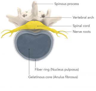
The bony parts of the spine, the vertebrae, are made up of a short cylindrical vertebral body and the vertebral arch that is connected to it. The vertebral body bears the weight of the body and the vertebral arch protects the spinal cord that runs through the spinal canal from the head to the pelvis. On the vertebral arch there are also the spinous and transverse processes that are the points of attachment of the ligaments and muscles. In the thoracic spine or middle back, each transverse process is joined to a rib by a small joint.

The discs sit between the vertebral bodies. The elasticity of the discs is what gives the spinal column its elasticity. The discs in the spine have a sort of gel cushion inside that enables them to absorb shocks. This watery gel-like core is surrounded by a ring of fibrous tissue. As you age, the elasticity of this cushion, and therefore your ability to move, and the size of the vertebral body decreases.

The spinal cord is the main nerve path in the human body. So that this important nerve pathway is well protected, it is located inside the spinal canal. Between the vertebral bodies, at about the level of the discs, branches of the spinal cord exit along the length of the spinal canal. These nerve fibres are called nerve roots and are the starting point of the nerves that reach throughout the entire human body. This is how the brain gets feedback from every part of our body and can control our body.

The cervical or upper spine is made up of seven vertebrae (C1 to C7) that are different in a number of ways to the rest of the spinal column. Apart from being considerably smaller than the vertebrae in the lumbar spine, the first six cervical vertebrae have extra openings in the bone in the transverse processes through which the arteries and veins run to supply blood to the brain and the vertebrae C2 to C6 also have a split spinous process. The first two cervical vertebrae are the most special, however. The first cervical vertebra is called the atlas and does not have a vertebral body but instead looks like a ring and is connected to the skull by a joint surface. It is in turn connected to the second cervical vertebra, the axis, by a joint surface rather than a disc. The axis does not have a vertebral body either, instead having a large spinal canal. There is a cone-shaped bony projection on the front of the axis called the odontoid process (odontoid = tooth-shaped) that faces upwards. It is connected with the front arch of the atlas by joint surfaces. The axis provides several attachment points for different ligaments that stabilise the skull and the spinal column. The atlas and the axis together form a functional unit that enables us to nod. The seventh cervical vertebra has a large spinous process that protrudes backwards. Unlike the other cervical vertebrae it is not split and can be felt through the skin.

The lumbar spine is made up of five vertebrae (L1 to L5). Compared to the other vertebrae in the spine they are larger and the flexibility of the spine decreases along the length of the lumbar spine. Its vertebrae have a bean-shaped outline and the load placed on them is the largest, which is why problems develop most often in the lumbar spine. At the level of the first or second lumbar vertebrae, the spinal cord spreads out into individual nerve roots, the cauda equina (Latin for horse’s tail).

The sacroiliac joints are the joints between the tailbone, the sacrum and the iliac bone. They connect the spinal column, which tapers down to the tailbone, with the pelvis and transfer weight and force to the legs. The range of motion of these joints is very limited, however, because of a strong ligament.

The typical double-S shape of the spinal column not only enables humans to walk upright but also ensures mechanical optimisation of forces thanks to the optimal positioning of the individual sections of the spine. This means that despite the heavy loads, hardly any effort is needed to stand upright, sit and hold the head upright. This optimal distribution of forces – the sagittal balance – is based on the many different angles and axes that are formed not only by the spine with its individual sections but also the pelvis. If this structure is pushed out of balance by any of a number of factors, the result is pain, degeneration or deformities.
However, all these ideal angles and axes are only theoretical textbook definitions and must in practice be determined individually for your spine. This is because every human has a unique anatomy, in the same way that we all have a unique personality.
So that parts of the body can be defined more accurately, they are divided into individual planes and axes. Just like in geometry, each plane is formed by two axes.
The medial plane divides the body into two equal halves, right and left. It is formed by the longitudinal or vertical axis that runs from the head to the feet or the reverse and the sagittal axis that runs from the front of the body to the back or the reverse.
The same axes also form the sagittal plane that runs parallel to the medial plane. It also divides the body into left and right sections. However, the sagittal plane is not necessarily in the middle of the body, which means that the two parts do not have to be equal.
The transverse or horizontal plane refers to all cross-sections of the body, regardless of whether the plane runs through the head, the middle of the body or the legs. It is formed by the sagittal axis that runs from the front to the back or the reverse and the transverse axis that runs from right to left or from left to right.
The frontal or coronal plane is parallel to the forehead and describes the front of the body. It is formed by the longitudinal and transverse axes.
In radiographic images of the body, that is, X-rays or CT or MRI scans, the planes referred to are the sagittal, the coronal, the axial and the AP view. A sagittal section shows a view from the side while a coronal section shows a view from the front. In an axial section, the body can be viewed in cross-section. In an AP image, the body is viewed from the front to the back or from the back to the front. This term is used mainly for X-ray images. Unlike CT or MRI scans, where several sections are imaged, with an X-ray only one image is taken in which all the organs are shown overlapping one another.
Patients
Latest News


