Request form
The US version of the website is under construction.
Important:
SIGNUS Medizintechnik GmbH only provides general information about spinal conditions. Please direct specific questions about your situation to your doctor. We cannot accept liability for incorrect indications and/or treatment and their consequences.
Imaging Procedures
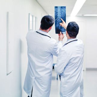
To ensure the right diagnosis, it is important that doctors can ‘look inside the patient’. This is possible using different imaging procedures such as X-rays, computed tomography (CT), magnetic resonance imaging (MRI) or sonography. These imaging procedures are needed not only for the diagnosis but also during surgery to precisely locate the area being operated on or during follow-up care to determine how successful the surgery has been. Along with a number of other procedures, X-rays and CT and MRI scans are important for spinal surgery.

The technique behind the image
X-ray diagnostics is a firmly established procedure in medicine that generates images of the internal organs and the skeleton. The procedure is based on the use of X-rays. These are electromagnetic waves with a wavelength between ultraviolet light and gamma radiation and are called ionising radiation because they can knock electrons out of atoms. X-ray radiation was discovered in 1895 by Wilhelm Conrad Röntgen in the Physics Institute of the University of Würzburg. Because of the ionising effect of X-rays, which means that they can damage cells, there must be a justifying indication for this examination for which the benefit outweighs the risk. Children and pregnant women in particular should avoid having X-ray examinations if possible.
So that X-ray radiation can be specifically used for medical diagnostics, the X-rays are generated in an X-ray tube. The high heating voltage at the cathode (negative pole) releases negatively charged electrons from the cathode material, which is usually tungsten. The high voltage between the cathode (negative pole) and the anode (positive pole) causes the electrons to accelerate rapidly towards the anode (positive pole). When the electrons hit the anode (positive pole) they are stopped abruptly, which releases energy known as the ‘braking radiation’ that is emitted in the form of X-rays. When the X-rays hit tissues in the body, they are taken up or absorbed to different degrees depending on the type of tissue. With a traditional X-ray machine, an X-ray film is placed opposite the X-ray tube that turns black when the X-rays hit it. The part of the patient’s body that is being examined is positioned between the X-ray tube and the film. Depending on how much radiation was absorbed by the body’s tissue, the film turns more or less black. This creates a negative picture of the tissue being examined. For example, bones, which absorb a lot of X-ray radiation, appear lighter than soft tissues that take up less radiation. Because the X-ray image is only a two-dimensional image, the organs being examined are shown overlapping one another. To be able to precisely identify the location of any abnormal features, X-ray images are therefore usually taken in at least two planes.
With a traditional X-ray image, the part of the body being examined is briefly irradiated with X-rays once and the resulting X-ray image therefore represents only a snapshot. In modern fluoroscopy, the X-ray image is not generated on X-ray film but the image is instead recorded digitally on a detector. This type of machine can maintain the X-ray radiation for longer and also record moving images. In addition, the parts of the body that strongly absorb radiation are shown in black, that is, the image is not a negative but rather a positive image. This procedure is used, for example, in an angiogram, an image of the blood vessels, or during surgery to enable implants to be positioned correctly.
Radiography is often used to examine bony structures or for initial diagnostics.
Computed tomography is used to image internal organs, again by using X-ray radiation. Unlike X-ray images, a CT scan does not produce a two-dimensional image but instead it generates cross-sectional images of the body. To be able to record individual cross-sectional images of the body, the gantry, a large ring that contains the X-ray tube and the detector on opposite sides to each other, rotates very quickly around the patient while the patient is continuously moved through the CT scanner. With each rotation, tomographic images are recorded from every angle around the patient, generating cross-sectional images like a ‘slice’ through the patient. The bed on which the patient lies is then moved a few millimetres further into the scanner so that the next ‘slice’ can be recorded. At the early days of computed tomography, the ‘slices’ were still very thick and between each ‘slice’ there were still layers of tissue that were not seen in the image. Since 1972 when the first CT scanner was developed, which had a fixed table position, there have been continuous improvements that have enabled the ‘slices’ to become thinner and thinner, and continuous recording is also now possible so that even the smallest abnormalities can be detected. Several sections can also now be recorded with each rotation and the use of several X-ray tubes means that the examination time is reduced further so that a whole-body scan can be carried out in just a few seconds. Another aim of recent improvements is to reduce the radiation dose even further.
Computed tomography enables bones as well as internal organs to be clearly imaged in cross-section. The very short examination time in particular means that it is possible to quickly obtain an overview of internal injuries and bleeding for trauma medicine.
Computer power also means that it is possible to generate three-dimensional simulations of the body using the two-dimensional cross-sectional images so that organs, bones or highly branched tumours can be better imaged.
Unlike X-rays and CT, no X-ray radiation or other ionising radiation is needed for magnetic resonance imaging but instead cross-sectional images are generated using a strong magnetic field and radio waves. No long-term complications are known for MRI, making it a gentle imaging procedure. The relatively long time needed for an examination of about 15 to 30 minutes and the noise made by the machine are the only drawbacks. It must also be determined before the examination that the patient does not have any ferromagnetic metals in or on the body, such as implants.
The principle behind nuclear magnetic resonance, another name for MRI, is nuclear spin. This is the property of protons, for example, a hydrogen proton, to spin around its own axis, which generates its own small magnetic field. In their natural state the protons of individual hydrogen atoms are not arranged in any order. If, however, they are placed in a strong magnetic field, such as that generated inside an MRI scanner, the protons either align themselves parallel or antiparallel to the magnetic field, like the needle in a compass. This process is called precession. The frequency with which the atomic nuclei precess around the longitudinal axis of the magnet is called the Larmor frequency and is about the same as radio frequency. Using a radio frequency pulse, which must correspond to the Larmor frequency, the hydrogen atoms are stimulated, which aligns them all in the same direction and brings them ‘into phase’. When the radio frequency pulse is switched off, the hydrogen atoms return to their previous state or they are said to relax.
There are two forms of relaxation that occur at the same time but independently of each other. T1 relaxation describes the increase in the longitudinal vector, that is, the time until about 63% of the protons are again aligned with the longitudinal axis. T2 relaxation describes the reduction in the transverse vector, that is, the time until only about 37% of the protons are aligned with the transverse axis.
The spin and therefore also the relaxation times of individual hydrogen protons depend on what types of bonds, that is, what type of tissue or liquid, they are in. Individual types of tissues have different T1 and T2 times which enables them to be displayed with different contrast in images. In a T1-weighted MRI image, the liquids are shown as dark and fat or high protein content is shown as light. In a T2-weighted image the liquids are light and fat is dark. Using these different images, the settings can be adjusted depending on the diagnostic aim or the tissue being examined.
MRI is well suited for imaging soft tissue or nerve fibres. Structures with a low water content such as bones or tissue containing lots of air such as lungs cannot be imaged so well, however.
Patients
Latest News


